Posts

Showing posts from July, 2018

Sleep Disturbances as a Risk Factor for Stroke

Image
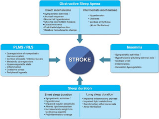
Sleep is an indispensable part of life, as with feeding and reproduction, all animal species require sleep. Humans sleep almost one-thirds of their lifetime,which is similar in industrialized and  in pre-industrial societies. Sleep, although characterized by quiescence and diminished responsiveness, is not a simple state of rest, but rather a cyclic state of periodic transitions between rapid-eye-movement (REM) and non-REM (NREM) sleep, which are precisely regulated by the central nervous system. Along with the brain and other organs or physiological streams, the cardiovascular system achieves homeostatic restoration during sleep, mainly through autonomic circulatory control. For example, the decrease in blood pressure during sleep, “dipping,” is a key biomarker of cardiovascular health, secondary to changes in activity and posture and also under the influence of sleep and circadian rhythms. During NREM sleep, the largest portion (up to 80%) of normal adult sleep, the autonomic

Time Is Brain: The Stroke Theory of Relativity

Image
Background Since the introduction of the philosophical tenet “ T ime is Brain! ,” multiple lines of research have demonstrated that other factors contribute to the degree of ischemic injury at any one point in time, and it is now clear that the therapeutic window of acute ischemic stroke is more protracted than it was first suspected. To define a more realistic relationship between time and the ischemic process, we used computational modeling to assess how these 2 variables are affected by collateral circulatory competence. Methods Starting from the premise that the expression “ Time = Brain ” is mathematically false, we reviewed the existing literature on the attributes of cerebral ischemia over time, with particular attention to relevant clinical parameters, and the effect of different variables, particularly collateral circulation, on the time–ischemia relationship. We used this information to construct a theoretical computational model and applied it to categorically d

Diagnostic Biomarkers for Stroke: A Stroke Neurologist’s Perspective

Image
Human stroke is remarkably heterogeneous. Stroke varies widely in severity with a majority of strokes being mild. Stroke comes in 3 major types. In North America and Europe, ischemic stroke comprises 85% of all strokes, intracerebral hemorrhage 8%, and subarachnoid hemorrhage 7%. These ratios differ in Asia with up to 30% of strokes occurring as intracerebral hemorrhages. For the neurologist, stroke is relatively easy to diagnose clinically. It is the most common cause of a sudden acute neurologic deficit in both adults and children. Imaging is the mainstay of identifying stroke type because it is not possible to distinguish ischemia from hemorrhage reliably on clinical grounds alone. A brain computed tomographic scan rules in an intracerebral hemorrhage or subarachnoid hemorrhage and in many cases rules in a diagnosis of ischemia. Acute computed tomography is insensitive to small-volume ischemia and therefore is usually normal in minor strokes and transient ischemic attack